Search


DNA Finger Printing
FINGER PRINTING:
DNA finger-printing (DNA typing, DNA identification, or genetic typing) is a technique involving chemically dividing the DNAinto fragments which form a unique pattern and then matching that “identity profile” with the pattern obtained from similarly testing a suspect’s blood specimen. If the two patterns match, the possibility of error, i.e. the chance that they do not belong to the same individual may be less than one in 30 billion. Dr. Alec Jeffreys in 1985, developed DNA fingerprinting.
Human body consists of about six thousand billion cells which constitute tissue and organ systems. Every living cell has genetic material contained in units called chromosomes, which are located in nucleus. DNA is present only in nucleated cells. Each human somatic cell has 23 pairs of chromosomes of which 23 are derived from biological father, and 23 from the mother, due to fertilization of ovum with sperm. Genes are arranged along the length of each chromosome, which are responsible for various functions of the body. Each gene carries instructions for the production of a particular protein which performs a particular function. Genes are also responsible for transmission of heredity. The genes are made up of chemical molecules called deoxyribonucleic acid (DNA). The human genome contains about 6x10 DNA molecules per diploid genome.
The core of the chromosome is a very long and extremely thin thread of DNA. A single human chromosome is about 1/12,500cm. long. The DNA molecule in this chromosome is about 2.5 cm. in length, compacted into the chromosome by successive coiling.
The total DNA in a cell is about 180 cm. in length. Each chromosome consists of two long linear DNA molecules, the polymers being hydrogen bonded via specific nucleotide pairing and coiled as a double helix which is spiral in nature, and looks like a spiral staircase. The helix is structurally stabilized by nuclear proteins called histones being referred to as chromatin. Chromatin may be condensed to varying degrees of compactness and in its most compact form is seen microscopically as chromosomes at the metaphase stage of each cell cycle. The chromosomes are continuous strands of DNA ranging from 50 to 500 million lolecules per chromosome, encoded in this.
Each nucleotide is composed of phosphate, deoxyribose sugar, and organic nitrogenous base. The bases are adenine (A), guanine (G), cystosine (C), and thymine (T). The bases of one strand are connected to the bases of the other strand b hydrogen bonds, while adjacent nucleotides are linked with each other by covalent bonds. Adenine combines only with thymine ad guanine combines only with cytosine. The DNA molecule resembles a twisted rope ladder with four kinds of stair-steps, e.g., A-T, T-A, C-G, or G-C. there are three hydrogen bonds between G and C, and 2 bonds between A and T.
A single DNA molecule consists of 50 to 500 million base pairs. The two strands of DNA helix run in opposite direction. The base sequence of one strand is always complementary to the sequence on the other. Each segment of DNA in a chromosome which codes for a particular protein is called a gene. In the human genome there are about 10,000 genes, accounting for about 5% of the entire cellular DNA. In between the active base pairs which code fro a particular protein, there are large number of redundant/inactive base pairs forming 95% of DNA, which is considered as “junk DNA”. In junk DNA short sequences of base, repeat themselves over again like a stutter (repetitive DNA), e.g. GCTA, GCTA, GATA, GATA, etc. The regions containing repetitive DNA demonstrating, hypervariability from person to person are called “satellite DNA”, which shows an extremely high degree of variability, and these variants are called “variable number tandem repeats” (VNTR) or “minisatellites”. there are more than 1500 VNTR’s in the human genome. Selected regions of VNTR are broken into fragments using special enzymes (restriction endonucleases), which are individualiatic in nature and establish 100% identity.
There are two methods of DNA analysis in common use. (1) RELP (restriction fragment length polymorphism). (2) PCR (polymerase chain reaction).
(1) RELP Method: DNA can be extracted from any body fluid or tissue in which nucleated cells are present. All the samples should be frozen semen, hair roots, tooth pulp, tissue from any organ, or skin) are usually examined. The separation of DNA involves. (1) distruption of cells and fractionation of cellular organelles, (2) dissociation of DNA from proteins by the use of salt solution or detergent, (3) addition of an extractant to phase-separate the bulk of the protein from the DNA, (4) use of enzymes or differential precipitation to remove the RNA and polysaccharides. The isoloated DNA is quantitated by ultraviolet spectrophotometry DNA is completely digested with restriction enzymes called restriction endonucleases. These enzymes recognize the specific sequence in the double strand DNA and cut the DNA at this site into various fragments, called as restriction fragment length polymorphism (RFLP). RFLP’s are produced due to variations in human DNA. These variations in restriction fragment lengths is due to presence of variable number of tandem repeats (VNTR). Most of repeated DNA are arranged as short sequences repeatedly contiguously in tandem, hence called VNTR. Several restriction rndonucleases are in use, e.g., Eco-R-I, PsT-1, HinF-I (obtained from E.coli), sau 3A-1 (obtained from Staphylococcus aureus), Hae-III (obtained from Haemophilus influenzae). PsT-1 (a six-base cutter) is commonly used, which recognizes the sequence CTGCAG.
Digested DNA is run on a agarose gel electrophoresis. The different restriction fragments are separated varying in length between 0.5 to 25 kb, which varies from one individual to another. The smaller fragments move much faster through the gel than the larger one. The gel is later stained with ethedium bromide for 40 minutes which tightly binds to DNA and fluoresces under UV light.
From the agarose gel, DNA is transferred to nylon membrane using capillary transfer technique of Southern. The result is a mirror-image replica of fragment distribution. Vacuum blotting of transfer is used more commonly as it is less time consuming. DNA is then fixed by heat at 800C or cross-linked by the cation of UV irradiation.
Next hybridization is done which is the pairing of two complementary single strands of DNA to form double stranded DNA. It involves the addition of a probe to the nylon membrane. A probe is a single-stranded recombinant DNA. Segment, or synthetic DNA, which is designed to go to a particular predetermined locus on a particular shromosome. It is usually tagged with a radioactive marker, such as P . The probe scans all the DNA fragments and wherever it encounters its complementary sequence, it will hybridise with it,thereby making the fragment radioactive. Usually four probes are used one at a time, due to which four different regions of the DNA would be alalysed.
In the Centre for DNA Fingerprinting and Diagnostics (CDFD), Nacharam, Hyderabad-76, A.P. India, Bkm probe is used, which is a multilocus probe isolated from the female banded krait, as a minor satellite DNA. Mor than 36 hypervariable VNTR are detected by BKm.
The label incorporated into the probe permits to localize these sites. After hybridization, the membranes are washed with 0.05% SDS which removes loosely bound probe. the membrane is then wrapped in the saran wrap and placed in the X-ray cassette holder along with X-ray film, and kept at minus 80 C. Exposure time depends upon the specific activity of the probe ranging from few hours to days (up to 10 days). X-ray films are then developed and fixed in the respective reagents and finally washed in water and dried. This autoradiograph is a permanent record, in which grey to black bands are seen where the radioactive probes had hybridized to fragments bearing complementary sequences. The series of bands seen on the film is called an “autorod”, which represents the DNA fingerprint of that individual from whom the DNA had been obtained. The pattern of bands is unique to each individual.
DNA FINGERPRINTING: All nucleated cells in the body contain 23 pairs of chromosomes. Each chromosome consists of a double spiral of DNA in the shape of a twisted ladder, the double helix. The DNA is composed of two strands of sugar and phosphate molecules that are twisted into a double helix that is bound together by links formed from adenine and thymine, and cytosine and guanine. Each helix twist has ten such links, like rungs on a twisted ladder. A single DNA molecule has thousands of such links, and the permutations of the bases in each adjacent link create the genes. The genes are separated by segments having no genetic function. Each human nucleus contains about a metre of DNA, but only 10% is used for genetic coding, the rest being redundant or silent segments (stutters; hypervariable regions (HVR); ministatellites). Of these redundant segment, there may be 200 to 14,000 repeats of each identical sequence on each DNA strand. These segments of nucleotides are called “repetitive DNA”. The length, constitution, and number of the repetitive sequences are different for each person, but are unique for an individual (except uniovular twins), and are transmitted from parents in regular fashion. This method is as unique as fingerprints to an individual.
Nucleated cells are the source of DNA for extraction from blood, semen, vaginal epithelial cells, tooth pulp, bone marrow, hair roots, muscle, skin, mucous membranes, etc. A sample is taken and from it the DNA is chemically extracted and purified. It is then cut into fragments at specific base sequences by a restriction enzyme (RE). When this process is repeated by several enzymes, each of which cuts at different sites, enough information is gathered to construct a detailed genetic fingerprint of a person. Because every person’s DNA specimen from one individual to another are different in number and length, from those in a DNA specimen from another individual. Next, the double stranded DNA are denatured into single strands. The DNA fragments are then separated by gel electrophoresis a negative charge and thus travels towards the positive pole in elctrophoresis. The distance that each fragment travels depends on its length. The longer the fragment, the slower its rate of migration. A number of DNA bands are produced in the gel. These are transferred to nylon or nitrocellulose sheets by Southern blotting technique. The membrane is then exposed to a DNA probe, which is a single strand of DNA labeled with radioactive phosphorus32, which specifically binds to the core sequences. X-ray film is put in direct contact with probe-labelled membrane to detect the radioactive patterns, which appear as a series of 30 to 40 parallel bands or bars. Each dark band represents a point where the DNA probe is bound to its complimentary base sequences. From the presence of different bars in given positions, comparisons can be made with other samples. It is possible to identify a person from a single human cell. The chance of two persons sharing the same sequence is about one in thirty thousand billions. Rarely, there are mutations confined to one band.
DNA analysis
1. From body of the diseased.
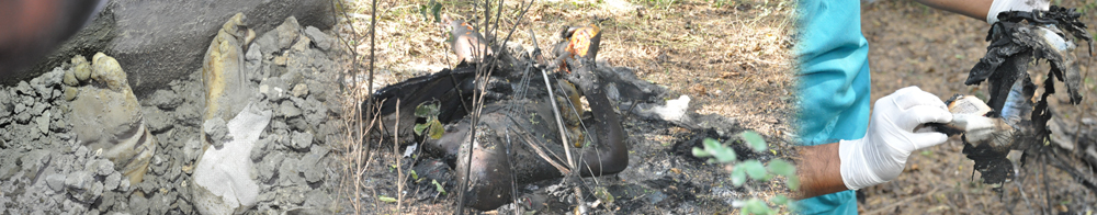
The biological samples should be collected in following priority order in case of dead person.
- Skeletal muscle ( deep muscle ) / Tissue – The concerned M. O. should collect the least decomposed portion of the tissue and it as soon as possible to avoid further decomposition.
-
Tough tissue – If the decomposition has already started collect the tougher
tissue for which the decomposition rate is comparatively slow.
Following types of tissue are observed to have lesser degree of decomposition.- Muscle tendons
- Foot i.e. heel skin
- Scalp skin
- Palm skin
- Stomach wall
- Tooth – Forword all the teeth present with the body.
- Scalp hair with roots – Forword bunch of scalp hair with roots. Pluck the scalp hair don’t cut them with scissors.
- P. M. Blood.
- Bones – If the skeletonisation of the deceased is complete and no tissue or other options are available, then the longer bones of the body such as Femur should be forworded. If some dried tissue or tendons are stuck up with the bones, then do not remove it, as there are some chances to get the DNA from it.
2. Crime scene and other Exhibits –
- Blood stains – In murder, attempt to murder and other related cases, blood stained clothes, scrapings, blood stained weapons and other related exhibits should be forwarded as per the requirement.
- Semen stains – In rape cases, clothes of victim and accused, clothes at crime scene, condoms and other related exhibits should be forwarded.
Mode of parcel and proper preservatives for respective samples.
|
Sr. No. |
Sample |
Mode of parcel |
Preservative |
|
01 |
Tissue, Muscle piece scalp skin etc. |
Put the sample in clean, sterile plastic or glass container add the preservative as recommended. Bring the sample in ice. |
DMSO or Normal physiological saline or 4% EDTA solution or keep the tissue as it is in –20 0 C refrigerator. |
|
02 |
Blood |
Clean sterile glass vial for P.M. blood. For control blood samples, tubes are supplied by FSL. Bring the samples in ice. |
4 % EDTA |
|
03 |
Teeth |
Air dry all the teeth available, put them in dry, clean and sterile plastic or glass container and forward. |
No preservative |
|
04 |
Scalp hair |
Air dry the sample, put in dry, clean and sterile plastic or glass container and forward. |
No preservative |
|
05 |
Bone |
Air dry and wrap in clean brown paper. Do not macerate or treat with any chemical. If the tissue or tendon is stuck upto the bone, keep as it is, do not separate or disturb it. |
No preservative |
|
06 |
Blood stained clothes and scrapings |
Air dry the clothes and wrap in clean brown paper. Don’t pack clothes in wet or semi wet condition. In case of blood stains on the wall, scrap with a clean new razor blade. While scraping, take the precaution that paint on the wall does not mix with the blood. Put the scrapings on clean piece of paper and prepare the packet. |
No preservative |
|
07 |
Semen stains |
Air dry the clothes and wrap in clean brown paper. Do not pack clothes in wet condition. In case of used condom, reverse it carefully to bring its inside out, empty it’s contents on a clean dry & sterile piece of cloth. Dry the cloth and forward. Also dry the condom pack in a brown paper and forward. |
No preservativ |